Meningiomas are tumors of the meninges, the membranes that surround and protect the brain and spinal cord. They are the most common type of primary brain tumor in adults, with most cases presenting as benign WHO Grade I. In contrast, high-grade meningiomas, which may be malignant, are less common and carry a higher likelihood of recurrence after resection. Although predominately benign, the high prevalence of this disease contributes to the growing medical burden. Furthermore, meningiomas can compress vessels and nerves and impinge on nearby brain tissue as they grow.
Meningiomas are highly vascular in nature, with growth, recurrence rate, and morbidity closely linked with neoangiogenesis, the process of formation of new blood vessels supporting the tumor microenvironment. Angiogenic factors that contribute to these pathways may be effective targets for therapies aimed at preventing tumor progression or lowering risk of recurrence. Currently, the standard care of treatment of meningiomas primarily comprises surgical resection with or without radiotherapy for high-grade tumors, yet prognosis remains poor. For benign cases, treatment often involves surgery and watchful waiting, which can often present a health burden for patients.
In close collaboration with Dr. Madhuri Wadehra’s lab at UCLA, we discovered increased mRNA expression of epithelial membrane protein 2 (EMP2) in meningioma. As EMP2 has been previously shown to promote neoangiogenesis in glioblastoma (GBM), we believe it holds a similar function in meningioma. These findings may support EMP2 as a potential molecular marker for angiogenesis in meningioma. Currently, our lab is investigating whether EMP2 is established in human-derived meningioma cell lines with baselines similar to what we see in primary tumors.
Most recently, our lab created a novel method of measuring hypervascularity through non-invasive neuroimaging to assess the benefit of preoperative embolization for intracranial meningiomas. Coined the Meningioma Vascularity Index (MVI), we present the MVI as a potential biomarker for meningiomas that may benefit from preoperative embolization.
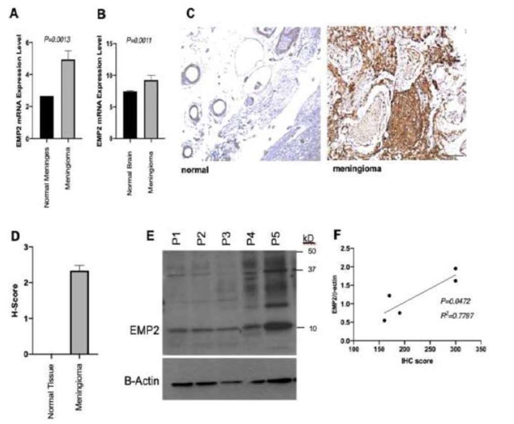
EMP2 mRNA expression is upregulated in meningioma speciments compared to regular meninges (A) and brain tissue samples (B). Immunohistochemistry straining with anti-EMP2 antibodies reveals an average of 95% positive staining among menigioma samples compared to regular brain speciments (C). Representative slides showing IHC staining with anti-EMP2 antibodies in meningeal and meningioma speciments (D). Parallel samples from a portion of the same tumor specimens were used for western blot analysis. Patel, K.S. et al. Identification of epithelial membrane protein 2 as a molecular marker and correlate for angiogenesis in mengioma. Journal of neuro-oncology J47.I (2020) 15-24 |
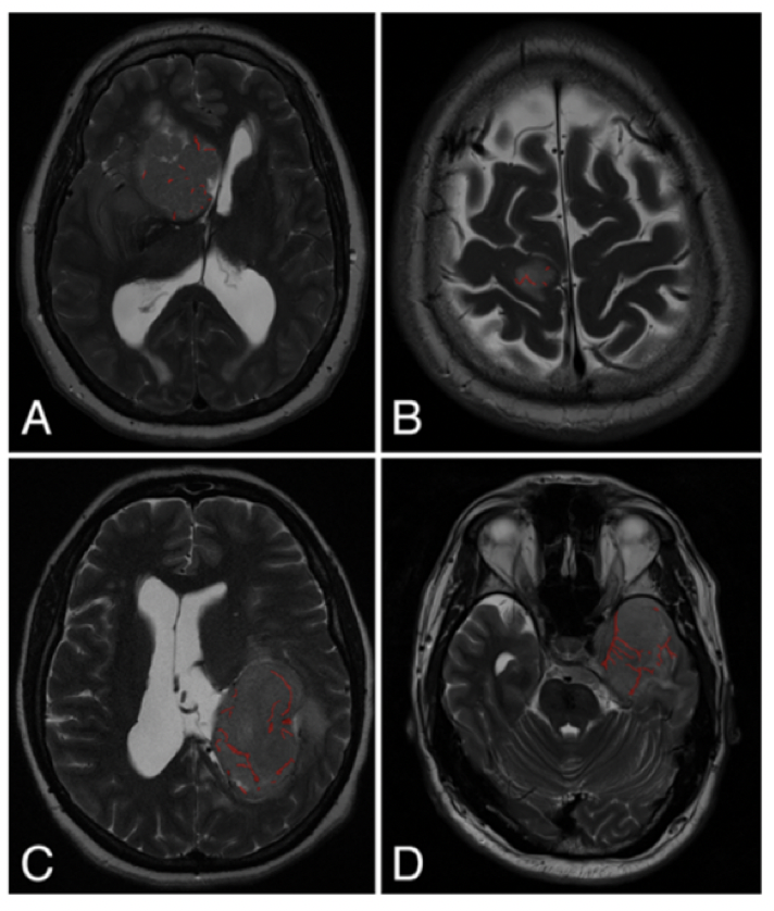
MVI Measured on axial T2-weighted MR images using the ITK-SNAP software. Lagman, Carlito et al. "The meningioma vascularity index: a volumetric analysis of flow voids to predict intraoperative blood loss in nonembolized meningiomas." Journal of neurosurgery J30.5 (2018): 1547-1552 |
2020
2018
2017